Wednesday 18 July 2012
Sound And Hearing
SOUND
• Sound is a type of energy that involves the vibration of
molecules in a medium, such as air or water.
• Sound is transmitted through a medium as a pressure wave.
• Sound vibrations are caused by an initial disturbance in a
medium that causes molecules around it to vibrate.
Examples: a guitar string being plucked, vocal chords
vibrating, vibrations of a diaphragm in a stereo speaker
• The rate at which sound vibrates is called the frequency of
the sound. Frequency is measured in Hertz (Hz).
• The amplitude of a sound wave is related to the loudness of
the sound. Loudness is measured in decibels.
HEARING
• Our ears receive and transmit sound wave vibrations to our
brains so that we can interpret the sounds around us.
• Humans can hear a range of frequencies from 20 to 20,000
Hz. Different animals can hear different ranges of sound
frequencies.
• Loud or high‐frequency sounds can cause damage to our
ears, causing hearing damage or hearing loss.
Credit:http://siemensscienceday.discoveryeducation.com/support-center/pdf/5-Minute_soundandhearing.pdf
Saturday 14 July 2012
Streoscopic Vision
Stereopsis (from stereo- meaning "solid" or "three-dimensional", and opsis meaning appearance or sight) is the impression of depth that is perceived when a scene is viewed with both eyes by someone with normal binocular vision.
Importantly, stereopsis is not usually present when viewing a scene with one eye, when viewing a picture of a scene with both eyes, or when someone with abnormal binocular vision (strabismus) views a scene with both eyes. This is despite the fact that in all these three cases humans can still perceive depth relations.
Example animal of streoscopic:owl
Friday 13 July 2012
Monocular Vision
Monocular vision is vision in which each eye is used separately. By using the eyes in this way, as opposed by binocular vision, the field of view is increased, while depth perception is limited. The eyes are usually positioned on opposite sides of the animal's head giving it the ability to see two objects at once.
Most birds and lizards (except chameleons) have monocular vision. Owls and other birds of prey are notable exceptions. Also many prey have monocular vision to see predators.
Credit: http://en.wikipedia.org/wiki/Monocular_vision
Most birds and lizards (except chameleons) have monocular vision. Owls and other birds of prey are notable exceptions. Also many prey have monocular vision to see predators.
Credit: http://en.wikipedia.org/wiki/Monocular_vision
Thursday 12 July 2012
Defects Of Vission (Astigmatism)
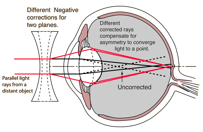
Astigmatism: This defect is when the light rays do not all come to a single focal point on the retina, instead some focus on the retina and some focus in front of or behind it. This is usually caused by a non-uniform curvature of the cornea. A typical symptom of astigmatism is if you are looking at a pattern of lines placed at various angles and the lines running in one direction appear sharp whilst those in other directions appear blurred. Astigmatism can usually be corrected by using a special spherical cylindrical lens; this is placed in the out-of-focus axis.
Defects Of Vission (Hyperopia)
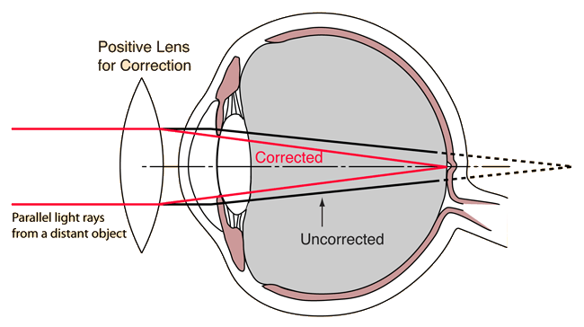
Hyperopia: (farsightedness) This is a defect of vision in which there is difficulty with near vision but far objects can be seen easily. The image is focused behind the retina rather than upon it. This occurs when the eyeball is too short or the refractive power of the lens is too weak. Hyperopia can be corrected by wearing glasses/contacts that contain convex lenses.
Credit:http://hyperphysics.phy-astr.gsu.edu/hbase/vision/eyedef.html and http://www.chm.bris.ac.uk/webprojects2002/upton/defects_of_the_eye.htm
Defects Of Vission (Myopia)
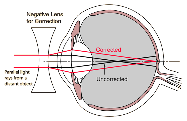
Myopia: (nearsightedness) This is a defect of vision in which far objects appear blurred but near objects are seen clearly. The image is focused in front of the retina rather than on it usually because the eyeball is too long or the refractive power of the eye’s lens too strong. Myopia can be corrected by wearing glasses/contacts with concave lenses these help to focus the image on the retina.
Wednesday 11 July 2012
Tuesday 10 July 2012
Focusing On Disntant And Near Object
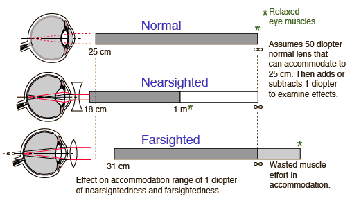
•Taking a nominal 50 diopter lens strength for the eye's lens combination, the effect of just a 1 diopter change in the lens strength upon accommodation can be assessed. It is more dramatic than you might expect with a 1 diopter nearsigntedness taking away clear vision beyond 1 meter and only adding 7 cm to the close-focusing capability. Fortunately, this defect can be corrected with just a -1 diopter corrective lens.
•The person with 1 diopter of far-sightedness can go for many years without knowing it because they can focus clearly to infinity, and with a nominal close-focus distance of 31 cm, young eyes can likely focus more closely with effort. But with the loss of pliability of the internal lens in later years, they may have eye-strain headaches and reach the point where they cannot read at normal distances.
Credit: http://hyperphysics.phy-astr.gsu.edu/hbase/vision/accom2.html
Monday 9 July 2012
Mechanism Of Sight
•The light rays from the object pass through the conjuctiva, cornea, aqueous humour, lens and vitreous humour in that order. All these structures refract the light such that it falls on the retina. This is called focussing. Maximum focussing is done by the cornea and the lens. The light then falls on the retina.
•This light is received by the photoreceptors - rods and cones, on the retina. The absorbed light activates the pigments present in the rods and cones. The pigments are present on the membranes of the vesicles. Thus, the light is then converted into action potentials in the membranes of the vesicles. These travel as nervous impulses through the rod or the cone cell and reach the synaptic knobs. From here the impulses are transmitted to the bipolar nerve cells, then to the ganglions and then to the optic nerves.
• Thus the nervous impulses generated in the retina are carried to the brain by about a million neurons of the optic nerve. The vision is controlled by the occipital lobe at the back of the brain. The information received is processed and we are able to see the image.
•The image formed on the retina is inverted. However, the brain makes us see the image erect. So, though the eyes are essential for vision, any damage to the optic nerves also results in impairment of vision.
The Structure Of Eye
•The eye is a complex optical instrument consisting of several parts.
•The cornea is exposed to the outside environment and therefore must repair itself rapidly because it is constantly faced with abrasion. It is transparent to all visible and near infrared wavelengths.
•The lens is transparent to visible and near infrared light. Together with the cornea, the lens focuses light to the back of the eye. The shape of the lens changes to accommodate near or distant viewing.
•The retina is the layer of nerve cells that receives the image and sends signals to the brain.
Credit: http://ehs.unc.edu/training/self_study/laser/eye.html
Saturday 7 July 2012
The Sense Of Hearing
• The ear is the sensory organ of sound.
• The sense of hearing is sensitive to the sound stimuli.
• The human ear can be divided into three main parts. These are known as the outer ear, the middle ear and the inner ear.
• Every structure of the ear has their own functions and are very important.
Outer Ear
Middle Ear
Inner Ear
Credit: http://pmr-science.wikispaces.com/1.5+The+Sense+of+Hearing
• The sense of hearing is sensitive to the sound stimuli.
• The human ear can be divided into three main parts. These are known as the outer ear, the middle ear and the inner ear.
• Every structure of the ear has their own functions and are very important.
CLICK THE PICTURE TO SEE MORE CLEARLY
Outer Ear
| Structure | Function/Explanation |
| Pinna | Made of cartilage and skin and shaped like a funnel. It collects and directs sounds into the ear canal. |
| Ear canal | A long tube lined with hairs. It directs sounds to the eardrum. |
Middle Ear
| Structure | Function/Explanation |
| Eardrum | A thin membrane that seperates the outer ear from the middle ear. It vibrates and transmits sound waves to the ossicles. |
| Ossicles | Made up of three small bones which is the hammer, the anvil and the stirrup. It intensifies the vibrations of the sound waves by 22 times before transmitting to the oval window. |
| Eustachian tube | A narrow tube that joins the middle ear to the throat that balances the air pressure at both sides of the eardrum. |
| Oval window | An oval-shaped, thin membrane between the middle ear and the inner ear. It transmits sound vibrations from the middle ear to the inner ear. |
Inner Ear
| Structure | Function/Explanation |
| Cochlea | Filled with liquid and contains the ends of nerve cells. The vibration of the oval window causes this liquid to vibrate. The vibration is detected by the nerve cells and are then changed into impulses. |
| Auditory nerve | It carries the impulses to the brain which then interprets the impulses as sound. |
| Semicircular canals | For body balance. |
Credit: http://pmr-science.wikispaces.com/1.5+The+Sense+of+Hearing
Friday 6 July 2012
The Sense Of Taste
Our tongue is the sensory organ for taste.
It can detect four basic tastes :
• Salty
• Sweet
• Sour
• Bitter
It can detect four basic tastes :
• Salty
• Sweet
• Sour
• Bitter
• The chemicals of the food dissolve in our saliva as we chew. The dissolved chemicals then stimulate the taste receptors in our taste buds to produce nerve impulses, which are then sent to the brain where they will be indentified as tastes.
Thursday 5 July 2012
The Sense Of Smell
●Our nose is a sensory organ of smell.
●The nostrils open into a hollow space called a nasal cavity.
●Mucous in the nasal cavity lines warms and moistens the air before it enters the lungs.
●The roof of the nasal cavity has many receptors and sensory cells to detect smell.
●Chemicals released by food, perfume and flowers into the air are known as smells.
●The chemicals dissolve in the mucous lining and stimulate the sensory cell which in turn, send out nerve impulses to the brain which interpret them as a smell.
CLICK THE PICTURE TO SEE MORE CLEARLY
●We get used to a smell after smelling something for a long time.This is because the sensory cells stop sending messages to the brain.The sensitivity of smell differs from individuals.
Wednesday 4 July 2012
The Sense Of Touch
Our skin consists of 3 main layers which are the epidermis, dermis, and the fatty layer.
Epidermis
- The epidermis is the outer layer of the skin, the epidermis itself is divided into three layers
- The outer most layer of the epidermis is the dead cell
The epidermis is a tough and water resistance layer that limits water loss
- Epidermis also protects the sensitive cells under it and prevents the entry of germs into your body
Dermis
- The dermis is the inner layer of your skin
There are glands, receptors and blood vessel in the dermis
- There are two glands in the dermis, which are sweat gland and sebaceous gland
- There are five receptors in the dermis, which are pain, pressure, heat, cold and touch receptors.
Touch receptors are sensitive to a slight pressure and are located at the top of the dermis.
Pain receptors are sensitive to pain.They are located at the top of the skin in the epidermis area to detect pain.
Heat receptors are sensitive to heat and is located in the dermis.
Cold receptors are sensitive to cold and is located in the dermis.
Pressure receptors detect pressure. For example, you feel pressure when carrying something heavy.Pressure receptors are located in the fatty layer deep into the skin.
When the receptors are stimulated, they send impulses to the brain.The brain interprates the impulses and sends them back along the nerves to the body to make a response.
Sumber: http://pmr-science.wikispaces.com/1.2+The+Sense+of+Touch
Subscribe to:
Posts (Atom)


